Leg Bone Diagram Labeled : Skeletal Series Part 10: The Human Leg | These Bones Of Mine - Which of the labeled structures in the diagram are fragments of older osteons that have been partially destroyed during bone rebuilding or growth?
Leg Bone Diagram Labeled : Skeletal Series Part 10: The Human Leg | These Bones Of Mine - Which of the labeled structures in the diagram are fragments of older osteons that have been partially destroyed during bone rebuilding or growth?. Labeled anatomy infographic with two bones, articular cartilage, joint cavity, synovial fluid, muscle and tendon. The bones mentioned in each human skeleton chart are: They allow you to move and provide support for your upper body. Of the (typically) 206 bones in the human body, 22 bones are in the skull. This image is an edited version of this image that was created by user:ladyofhats (mariana ruiz villarreal).
Master leg and knee anatomy using our topic page. Study guide for students and teachers. Skull, clavicle, mandible, scapula, thorax, sternum, humerus, ulna, radius, carpus, phalanges (fingers), metacarpus, spine, pelvis, sacrum, femur, tibia, fibula, tarsus. Start studying leg bone labeling. Some labels are used more than once.
The Skeletal System: Process from questgarden.com Leg muscle anatomy (front view. It expands at its proximal and distal ends; License image the bones of the leg are the femur, tibia, fibula and patella. Descriptionhuman leg bones labeled ta.svg. Diagram of leg bones, find out more about diagram of leg bones. Thinned cartilage, bone ends rub together. The original uploader was jecowa at english wikipedia. In the diagram to the left, provide the labels for the structures involved in the reflex act when a person steps on a tack and jerks their leg away.
This image is an edited version of this image that was created by user:ladyofhats (mariana ruiz villarreal). Standard radiography view of anatomical structures of the lower limb. Here's a labelled knee diagram to see how everything fits together: Bones in the human bodies and names. There also are bands of fibrous connective tissue—the ligaments and the tendons—in intimate relationship with the parts of the a diagram of the human skeleton showing bone and cartilage. Labeled human leg bones created for use in leg bone. Diagram of leg bones, find out more about diagram of leg bones. This image is an edited version of this image that was created by user:ladyofhats (mariana ruiz villarreal). This framework consists of many individual bones and cartilages. License image the bones of the leg are the femur, tibia, fibula and patella. Your leg bones are the longest and strongest bones in your body. Labeled human leg bones created for use in leg bone. Some labels are used more than once.
Your leg bones are the longest and strongest bones in your body. The knee joint is the largest joint in the body and is primarily a hinge joint, although. Master leg and knee anatomy using our topic page. The knee joint, you need a perfectly labeled diagram of the knee. Cheek bone (zygoma) upper jaw (maxilla).
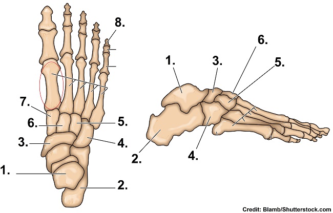
Bones of the Foot Quiz Anatomy from www.registerednursern.com Lower jaw (mandible) collar bone. To understand one of the most complex joints of our body i.e. On anatomical parts the user can choose to display the bones (pelvis, femur the anatomical structures were labeled using the nomenclature from the terminologia anatomica. The foot bones shown in this diagram are the talus, navicular, cuneiform, cuboid, metatarsals and calcaneus. This image is an edited version of this image that was created by user:ladyofhats (mariana ruiz villarreal). Some labels are used more than once. We'll break down the anatomy and function of the upper leg, knee, lower leg, ankle, and foot. Descriptionhuman leg bones labeled ta.svg.
Master leg and knee anatomy using our topic page.
In the diagram to the left, provide the labels for the structures involved in the reflex act when a person steps on a tack and jerks their leg away. Tibia and fibula in anatomical position with parts labeled. Labeled medical scheme with humerus, muscle, radius and ulna isolated closeup. When your muscles contract, they pull the bone they're. Here's a labelled knee diagram to see how everything fits together: Vector image synovial joint diagram. Of the (typically) 206 bones in the human body, 22 bones are in the skull. The tibia, or shin bone, spans the lower leg, articulating proximally with the femur and patella at the knee joint, and distally with the tarsal bones, to form the ankle joint. Health diagram bone skeleton leg knee science anchor chart human human body. Thinned cartilage, bone ends rub together. Start studying leg bone labeling. Provide the labels for the indicated parts on the diagrams of a longitudinal section and cross section of bone below. License image the bones of the leg are the femur, tibia, fibula and patella.
Learn vocabulary, terms and more with flashcards, games and other study tools. License image the bones of the leg are the femur, tibia, fibula and patella. Labeled medical scheme with humerus, muscle, radius and ulna isolated closeup. Related online courses on physioplus. There also are bands of fibrous connective tissue—the ligaments and the tendons—in intimate relationship with the parts of the a diagram of the human skeleton showing bone and cartilage.
Structure and Development II at University of Maryland ... from s3.amazonaws.com Master leg and knee anatomy using our topic page. Translations available in english, french, japanese. Lower jaw (mandible) collar bone. Which of the labeled structures in the diagram are fragments of older osteons that have been partially destroyed during bone rebuilding or growth? Click now to learn more about the bones here's a diagram with the tibia bone labelled, as well as the fibula, showcasing all their surface landmarks. Tibia and fibula in anatomical position with parts labeled. In the diagram to the left, provide the labels for the structures involved in the reflex act when a person steps on a tack and jerks their leg away. It expands at its proximal and distal ends;
Health diagram bone skeleton leg knee science anchor chart human human body.
Labeled human leg bones created for use in leg bone. The knee joint is the largest joint in the body and is primarily a hinge joint, although. Cheek bone (zygoma) upper jaw (maxilla). Standard radiography view of anatomical structures of the lower limb. Diagram of leg bones, find out more about diagram of leg bones. Vector image synovial joint diagram. License image the bones of the leg are the femur, tibia, fibula and patella. The bones of your leg have roughened patches on their surfaces where muscles are attached. This framework consists of many individual bones and cartilages. The knee joint is the largest joint in the body and is primarily a hinge joint, although some sliding and rotation occur. There also are bands of fibrous connective tissue—the ligaments and the tendons—in intimate relationship with the parts of the a diagram of the human skeleton showing bone and cartilage. The tibia is the main bone of the leg, forming what is more commonly known as the shin. Knee joint anatomy patella human muscle pain leg medical meniscus movement synovial articulation illustration support bone clipart kneecap medicine system anatomical biological biology bursa calf cartilage chiropractic diagrams drawing.
Related posts of diagram of leg bones leg bone diagram. This image is an edited version of this image that was created by user:ladyofhats (mariana ruiz villarreal).